English
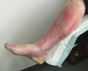
Discolouration of the skin in the ankle region occurs because of red blood cells and proteins leaking out of the capillary walls and into the interstitial fluid. The haemoglobin is then broken down outside the capillaries into the tissue, presenting as a visible brown colour called haemosiderin.
One should observe if the foot is dry and scaly as this can be a sign of venous insufficiency.
Patients with venous leg ulcers can develop varicose eczema where the surrounding skin is inflamed. Cell division occurs and white blood cells are drawn to the wound. The eczema may be as a result of contact dermatitis caused by the bandaging, stasis dermatitis, allergic contact dermatitis or irritative dermatitis due to the exudates irritating the skin on the leg.
Common sensitizers include wool alcohols (lanolin), topical antibiotics, topical corticosteroids, cetyl stearyl alcohols, parabens and rubber mixes.
Emollients are the first line treatment for varicous eczema. Emollients used as a soap substitute to wash and moisturise the leg is recommended. Topical corticosteroids ointments may also help control severe varicous eczema. These should only be used for a limited period of time as they only treat the symptoms of varicose eczema and long-term use can mask any underlying skin/leg infection.
8.2.3 Changes in the skin in relation to venous insufficiency