English
4.0 Classification of wounds 
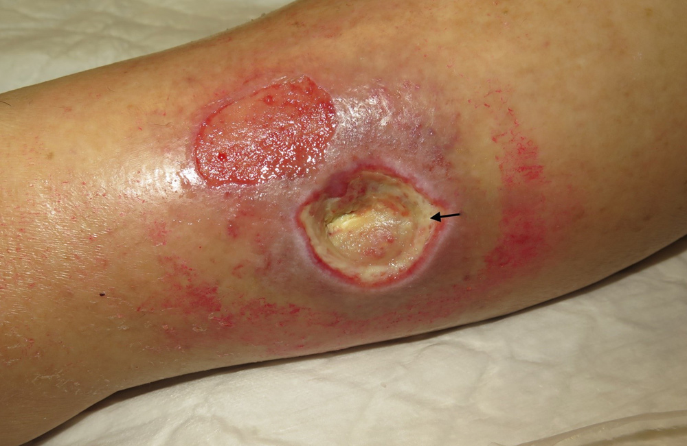
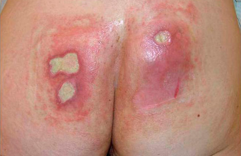
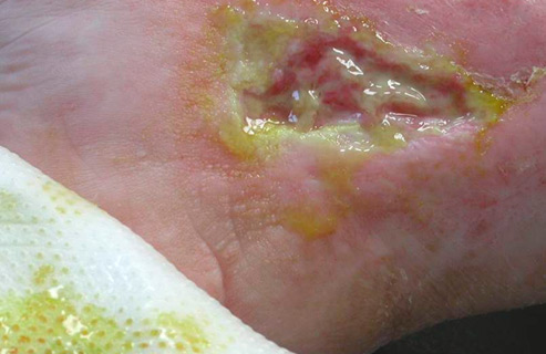
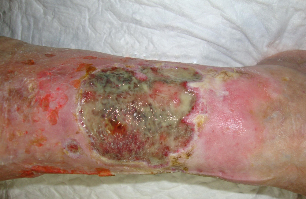
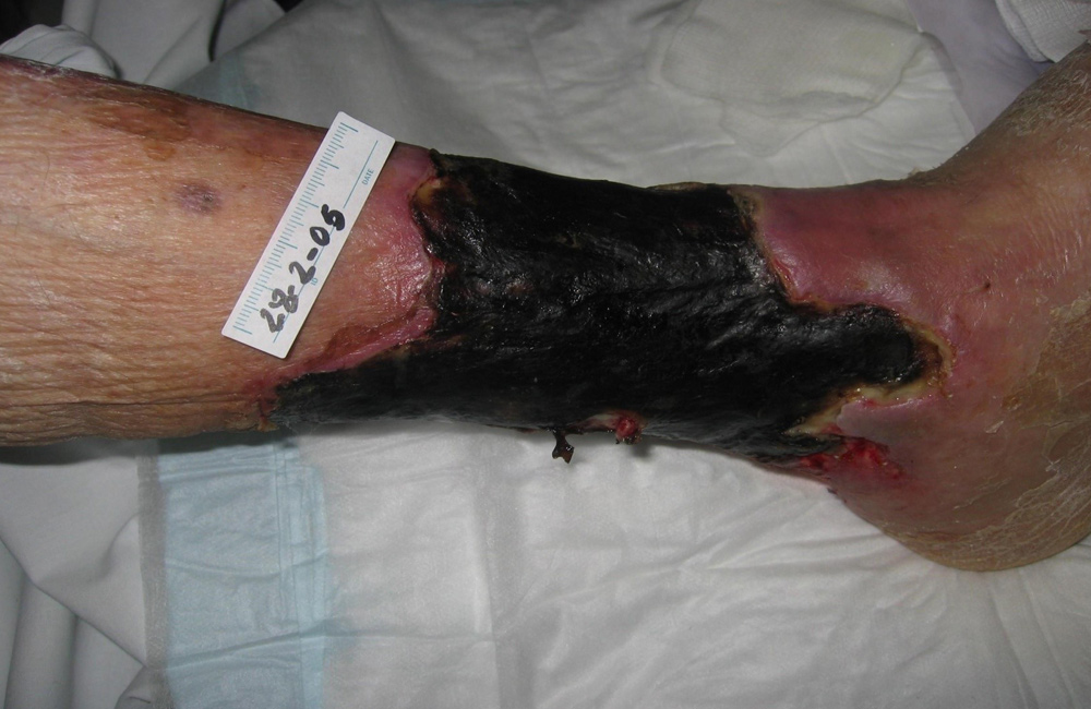
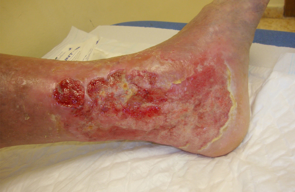
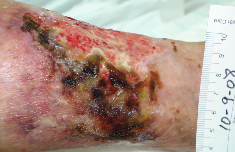
A wound can be defined as a breakdown in the protective function of the skin and can be divided into acute and chronic wounds. Acute wounds tend to heal in an uncomplicated way following the normal stages of wound healing. Chronic wounds tend to be full thickness and because the process of healing has become disrupted they take longer to heal. Taking longer to heal puts these chronic wounds at risk of becoming infected.
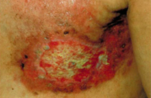
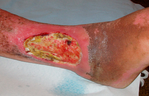
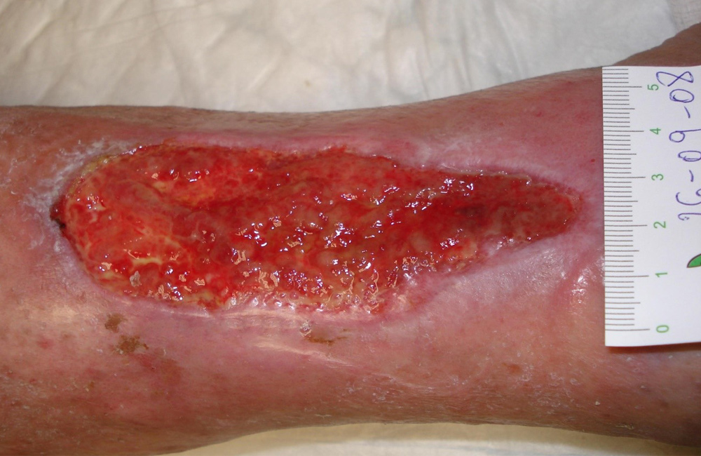
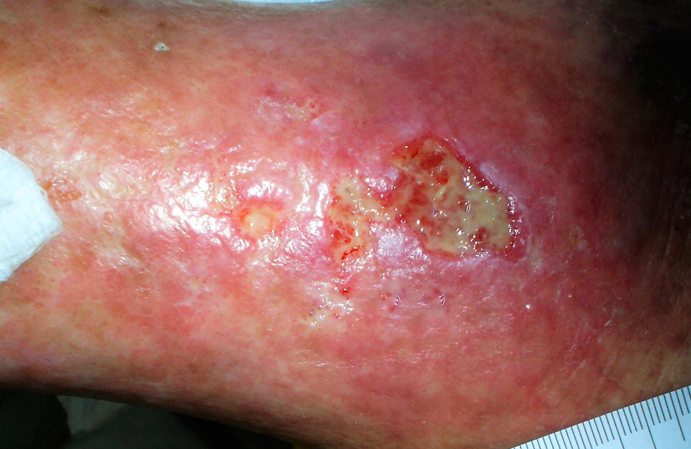
Both acute and chronic wounds differ in appearance (morphology) and causes (aetiology). Both wounds can appear as follows –
Appearance (morphology)
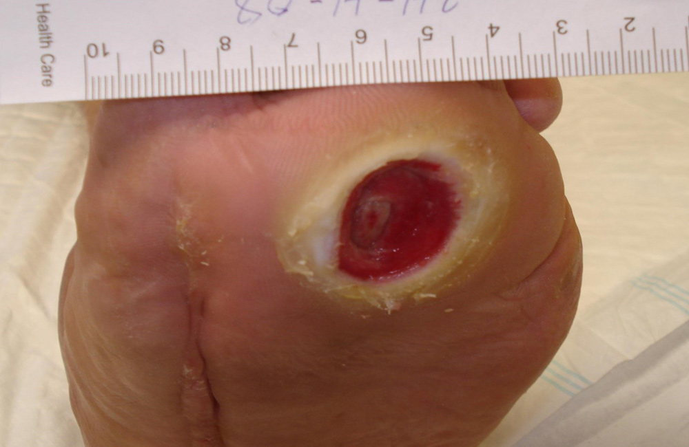
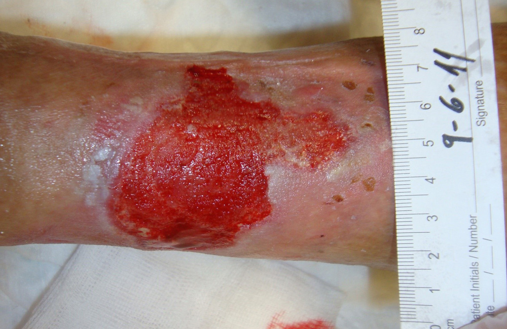
- Clean granulating wounds
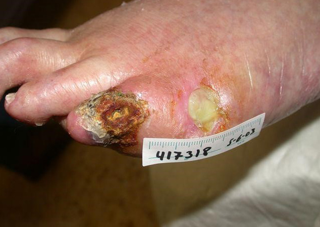
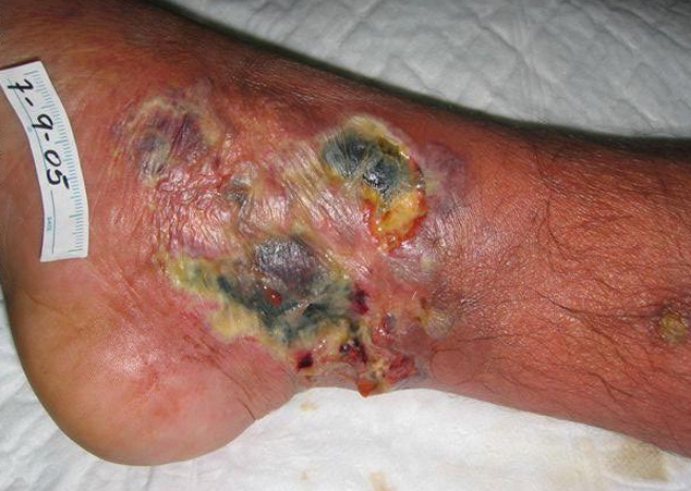
- Wounds with necrotic tissue
- Epithelialising wounds
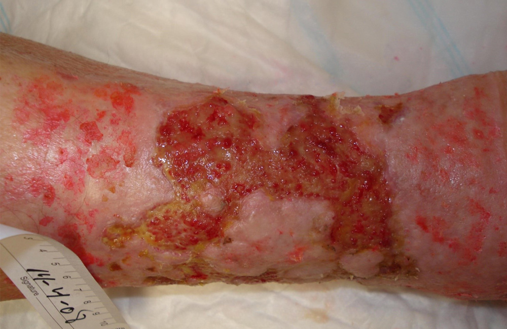
- Sloughy wounds
- Wounds with hyper granulation
- Wounds with excessive exudate
- Wounds where the granulation looks dull
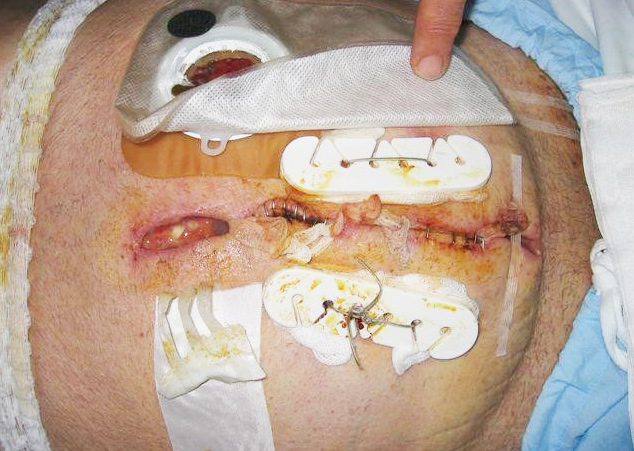
- Surgical wounds
Cause (aetiology)
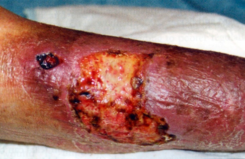
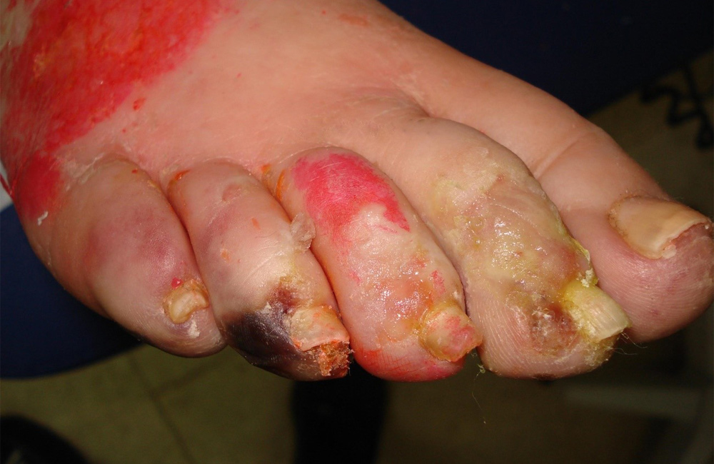
- Ulcers can be related to changes in vessels. It may be arterial, venous or lymphatic disease that has caused the ulceration
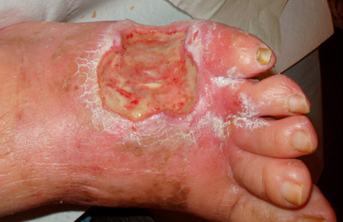
- Neuropathic ulcers related to diabetes
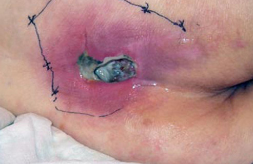
- Surgical wounds
- Burns
- Trauma wounds
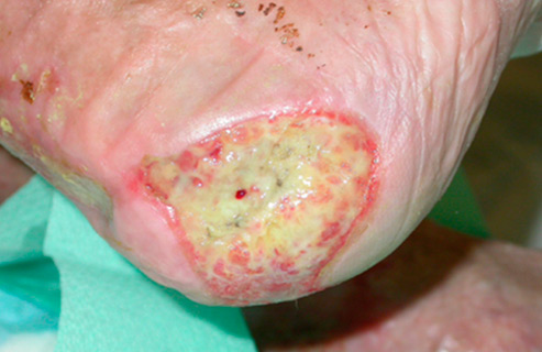
- Pressure ulcers
- Fungating tumours
- Radiotherapy burns
- Fistulas
- Dermatological cause
- Self-inflicted wounds