4.0 Classification of wounds Listen

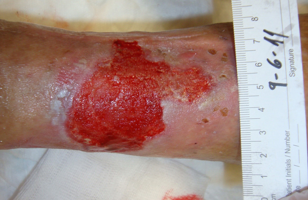
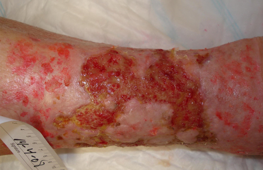
A wound can be defined as a breakdown in the protective function of the skin and can be divided into acute and chronic wounds. Acute wounds tend to heal in an uncomplicated way following the normal stages of wound healing. Chronic wounds tend to be full thickness and because the process of healing has become disrupted they take longer to heal. Taking longer to heal puts these chronic wounds at risk of becoming infected.

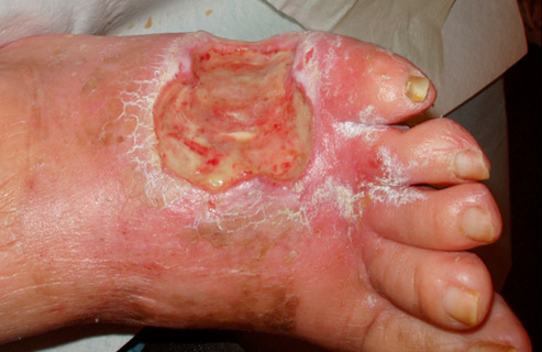
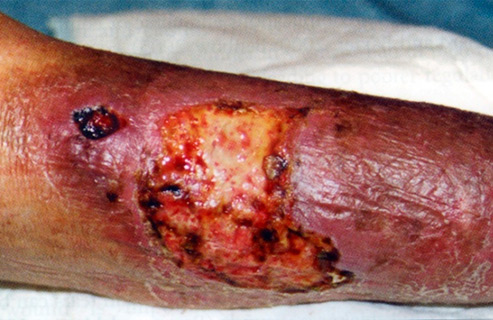
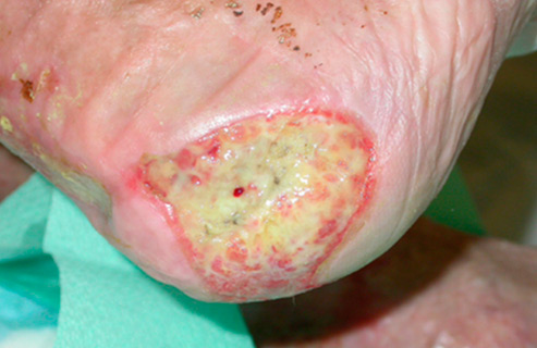
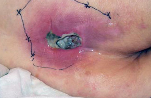
Both acute and chronic wounds differ in appearance (morphology) and causes (aetiology). Both wounds can appear as follows –

Appearance (morphology)

  • Clean granulating wounds
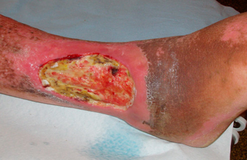
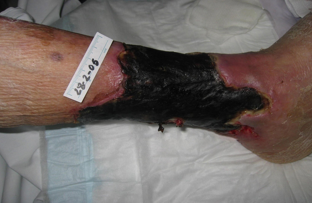
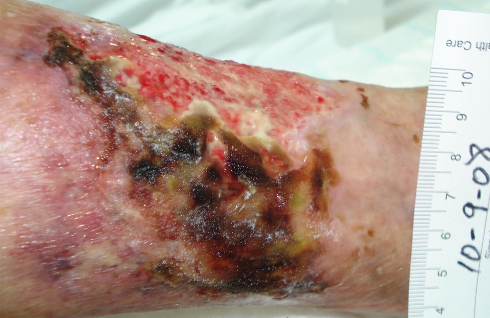
  • Wounds with necrotic tissue
  • Epithelialising wounds
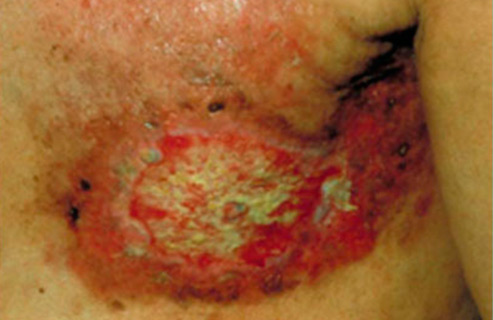
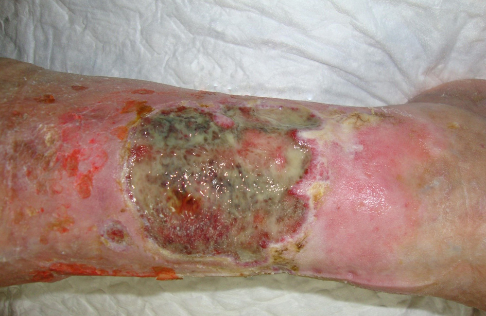
  • Sloughy wounds
  • Wounds with hyper granulation
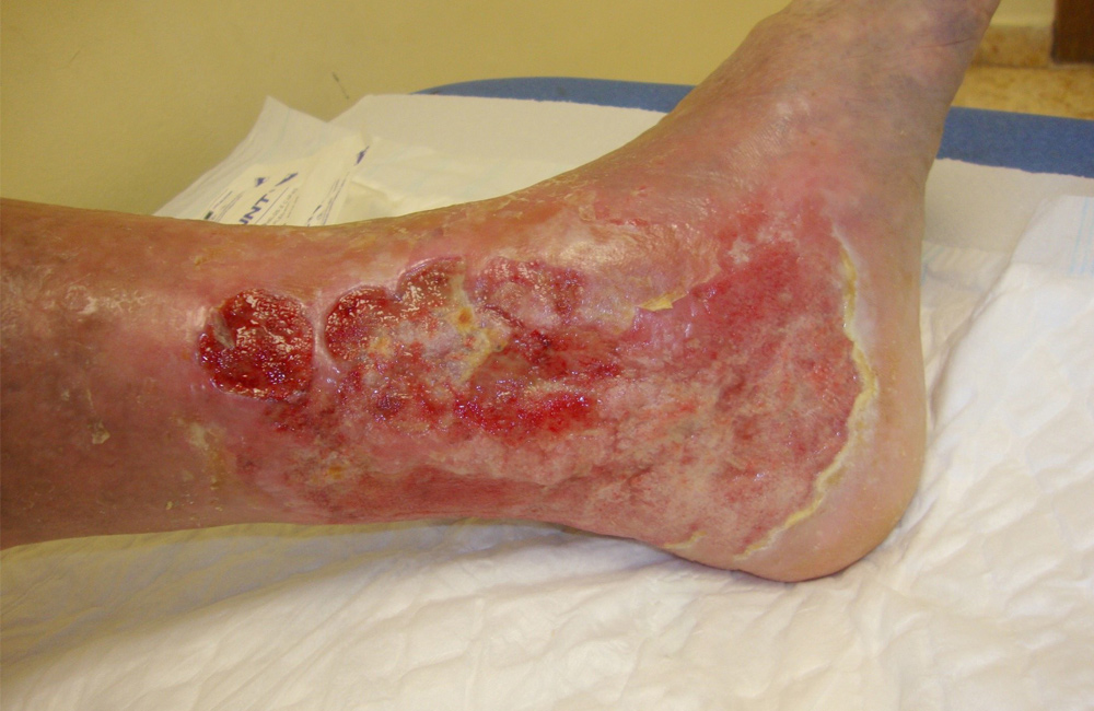
  • Wounds with excessive exudate
  • Wounds where the granulation looks dull
  • Surgical wounds

Cause (aetiology)

  • Ulcers can be related to changes in vessels. It may be arterial, venous or lymphatic disease that has caused the ulceration
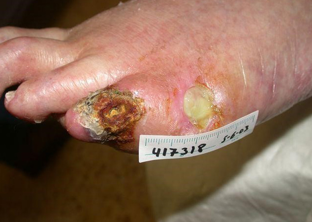
  • Neuropathic ulcers related to diabetes
  • Surgical wounds
  • Burns
  • Trauma wounds
  • Pressure ulcers
  • Fungating tumours
  • Radiotherapy burns
  • Fistulas
  • Dermatological cause
  • Self-inflicted wounds
 

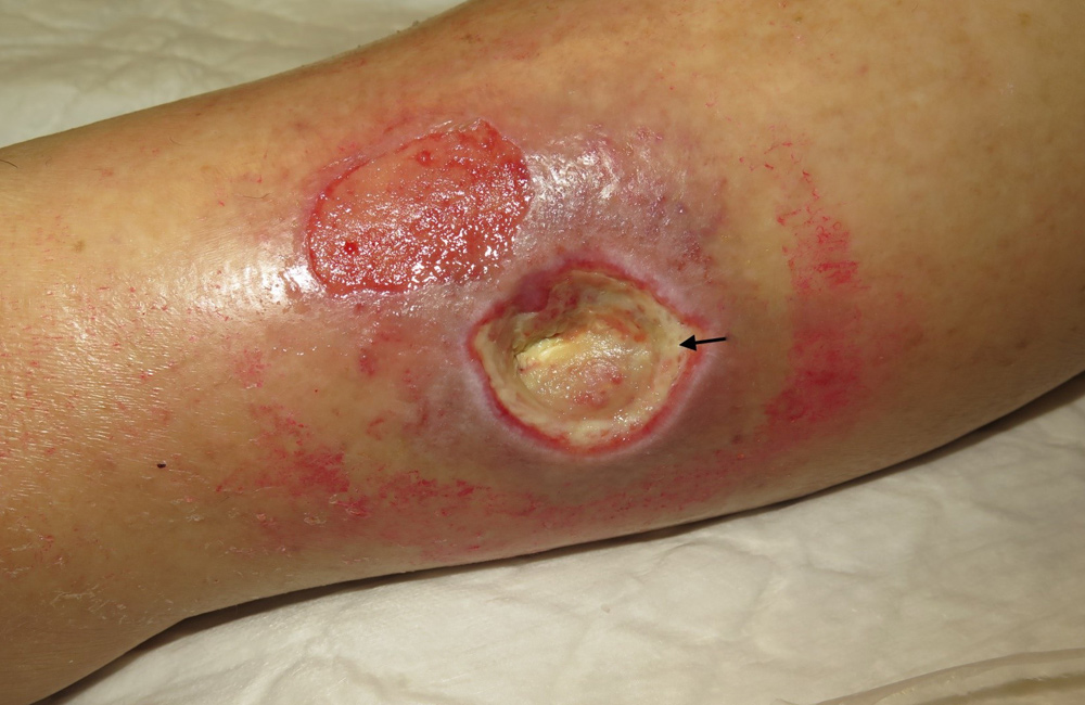
woundBedFibrin_1000x650px
Wound bed with fibrin coating
2_saar_nekrotisk
Wound with necrotic tissue ( yellowed necrosis)
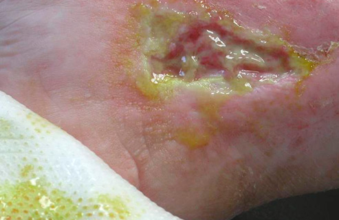
4_bakteriell_ubalanse
Wound with bacterial imbalance
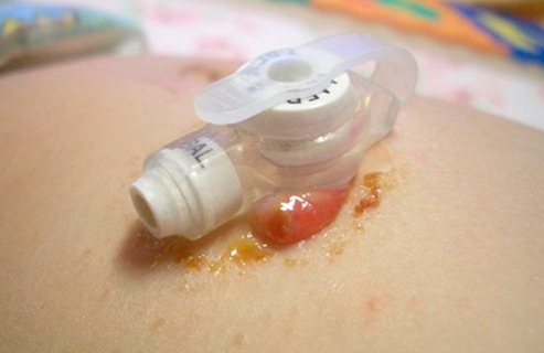
7_hypergranulasjon
Wound with hypergranulation tissue
6_dårligokysgenerte
Arterial wounds: can be due to blood vessel disorders related to arterial, venous or lymphatic problems
12_traumeskade
Trauma injuries: injuries, operations and heat related wounds are acute trauma wounds
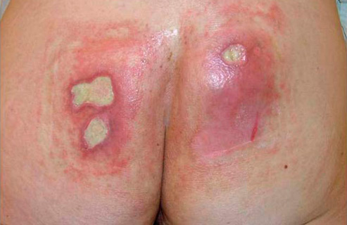
13_trykksaar
Pressure wounds: wounds caused by pressure and friction
14_trykksaar2
Pressure wounds: pressure wound with blackening and yellowing necrosis
15_kreftsaar
Cancer wound
16_straleskadet
Radiation injury to the skin
granulation_tissue1000px650
Granulation tissue
arterial_ulcer_1000px650
Arterial ulcer
bacterialbiofilm1000px650
Bacterial biofilm
dry_necrosis_1000px650
Dry necrosis
exudative_ulcer1000px650
Exudative ulcer
hypertensive_ulcer_1000px650
Hypertensive ulcer
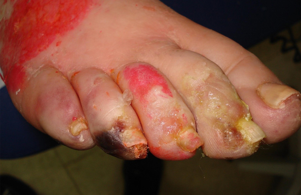
ischemia_in_diabetic_foot1000px650
Ischemia in a diabetic patient
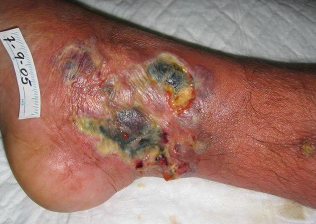
ischemic_ulcer
Ischemic ulcer
necrotising_fascitis_1000px650
Necrotising fascitis
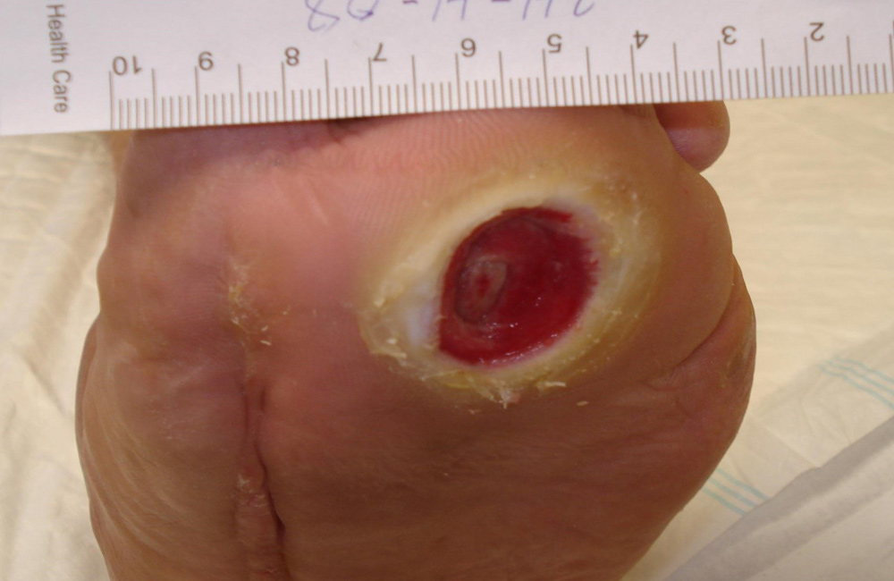
neuropathic_ulcer_in_diabetic_foot1000px650
Neuropathic ulcer in a diabetic foot
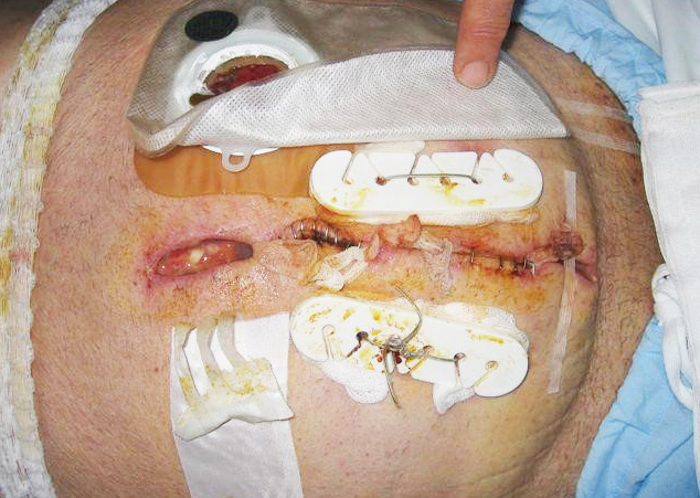
surgical_wound_
Surgical wound
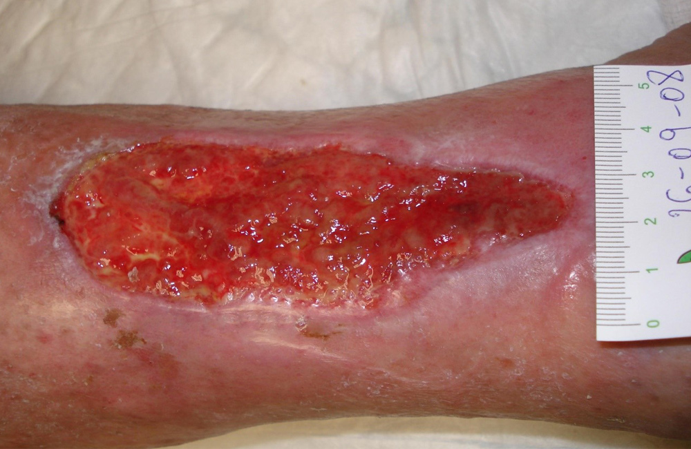
venous_ulcer1000px650
Venous ulcer I
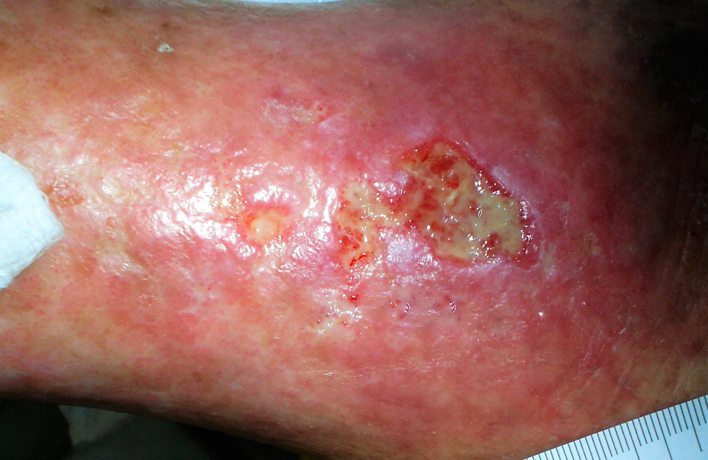
venous_ulcer_example2_1000px650
Venous ulcer II